What are the Ehlers-Danlos syndromes and hypermobility spectrum disorder?
What does EDS stand for?
The Ehlers-Danlos syndromes (EDS) are a group of thirteen individual genetic conditions, all of which affect the body’s connective tissue. Connective tissue is the body’s “support tissue”. It helps hold joints, skin and organs in place.
What does HSD stand for?
HSD stands for hypermobility spectrum disorder. Hypermobility means some joints bend further than usual. HSD is used when that extra movement is linked with symptoms, such as pain or joints that feel unstable.
Why we say EDS and HSD together
We write Ehlers-Danlos Syndromes and hypermobility spectrum disorder (EDS and HSD). We do this because:
- We support people affected by both
- Symptoms and day-to-day needs can be similar
- Some people are diagnosed with EDS, and some are diagnosed with HSD, but both groups might need the same kind of support
EDS and HSD symptoms can be mild or severe, and they can change over time. If they are affecting your daily life, it is a good idea to ask for support and advice.
What are the main symptoms of EDS and HSD?
People with EDS and HSD can have a wide mix of symptoms. You might have some of these, and not others.
If you have not had a diagnosis yet, you can use our Healthcare Professionals Hub at your first GP appointment to support the process. If you feel seriously unwell or unsafe, call NHS 111 or 999.
Types of EDS
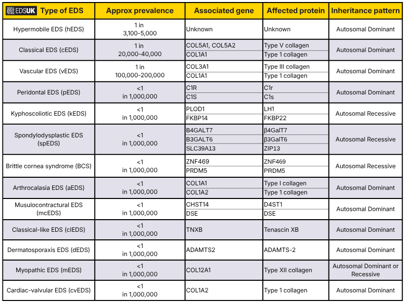
The Ehlers-Danlos syndromes (EDS) are split into 13 types. Most types are rare. Each type is its own condition and it is caused by a change in a specific gene. This means:
- One type does not turn into another type
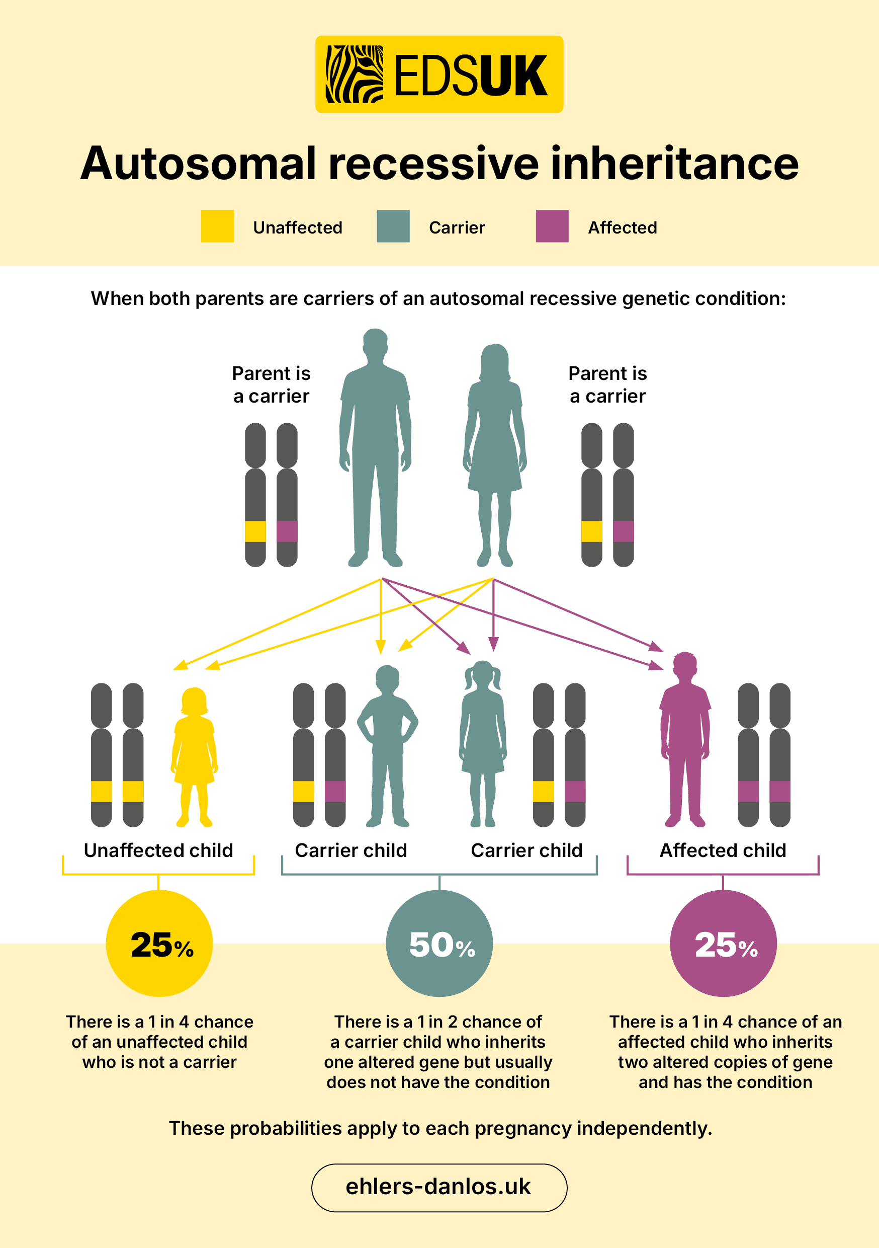
- A child cannot inherit a different type of EDS to the one their parent has (see poster below)
Some rare types can be confirmed with genetic testing. This is a lab test that looks for changes in your genes.
The most common type hypermobile EDS (hEDS) is treated and managed in the same way as hypermobility spectrum disorders and cannot be diagnosed currently with a genetic test. A GP would need to provide a clinical diagnosis.
These are the 13 types:
For more detailed information see the chart at the bottom of the page. Please note that the criteria may possibly be changing for some types in December 2026.
Arthrochalasia EDS (aEDS)
Please note: The following text cannot and should not replace advice from the patient’s healthcare professional(s). Any person who experiences symptoms or feels that something may be wrong should seek individual, professional help for evaluation and/or treatment. This information is for guidance only and is not intended to provide individual medical advice.
What is aEDS?
aEDS is an incredibly rare type of EDS and symptoms include severe joint hypermobility, hyperextensible skin, mild dysmorphic features, and bilateral congenital hip dislocation.
What is the cause of aEDS?
aEDS is caused by variations in a person’s genes. aEDS is caused by variations in genes COL1A1 or COL1A2.
How is aEDS diagnosed?
When diagnosing aEDS a list of common and relevant signs and symptoms of the condition are used. This is referred to as diagnostic criteria and in the case of aEDS it is split into a set of major and minor criteria.
The Major criteria
- Congenital bilateral hip dislocation
- Severe generalised joint hypermobility, with multiple dislocations/subluxations
- Skin hyperextensibility
The Minor criteria
- Muscle hypotonia
- Kyphoscoliosis
- Radiologically mild osteopenia
- Tissue fragility, including atrophic scars
- Easy bruisable skin
To meet the diagnostic criteria for aEDS a person must have:
Major criteria 1 (congenital bilateral hip dislocation)
plus
Major criterion 3 (skin hyperextensibility) Or major criterion 2 (severe GJH with multiple dislocations/subluxations) and at least two other minor criteria
If a person meets the criteria a genetic test is done to confirm the diagnosis.
How is aEDS managed?
aEDS is managed through addressing the symptoms a person is experiencing. This is best done through being in the care of the NHS National EDS service; a specialised service for adults and children who have or are suspected to have a rare type of EDS.
For information about the service and for referral eligibility please click on the following links:
EDS National Diagnostic Service (based in London) covering the South of England
EDS National Diagnostic Service (based in Sheffield) covering the North of England
Brittle cornea syndrome (BCS)
Please note: The following text cannot and should not replace advice from the patient’s healthcare professional(s). Any person who experiences symptoms or feels that something may be wrong should seek individual, professional help for evaluation and/or treatment. This information is for guidance only and is not intended to provide individual medical advice.
What is BCS?
BCS is an incredibly rare type of EDS and causes individuals cornea’s to be thin and fragile.
What is the cause of BCS?
BCS is caused by variations in a person’s genes. BCS is caused by variations in genes ZNF469 or PRDM5.
How is BCS diagnosed?
When diagnosing BCS a list of common and relevant signs and symptoms of the condition are used. This is referred to as diagnostic criteria and in the case of BCS it is split into a set of major and minor criteria.
The Major criteria
- Thin cornea, with or without rupture
- Early onset progressive keratoconus
- Early onset progressive keratoglobus
- Blue sclerae
The Minor criteria
- Enucleation or corneal scarring as a result of previous rupture
- Progressive loss of corneal stromal depth, especially in central cornea
- High myopia, with normal or moderately increased axial length
- Retinal detachment
- Deafness, often with mixed conductive and sensorineural components, progressive, higher frequencies often more severely affected (“sloping” pure tone audiogram),
- Hypercompliant tympanic membranes
- Developmental dysplasia of the hip
- Hypotonia in infancy, usually mild if present
- Scoliosis
- Arachnodactyly
- Hypermobility of distal joints
- Pes planus, hallux valgus
- Mild contractures of fingers (especially 5th)
- Soft, velvety skin, translucent skin
To meet the diagnostic criteria for BCS a person must have:
Major criteria 1 (thin cornea, with or without rupture)
plus
Either: at least one other major criterion
And/or three other minor criteria
If a person meets the criteria a genetic test is done to confirm the diagnosis.
How is BCS managed?
BCS is managed through addressing the symptoms a person is experiencing. This is best done through being in the care of the NHS National EDS service; a specialised service for adults and children who have or are suspected to have a rare type of EDS.
For information about the service and for referral eligibility please click on the following links:
EDS National Diagnostic Service (based in London) covering the South of England
EDS National Diagnostic Service (based in Sheffield) covering the North of England
Cardiac-valvular EDS (cvEDS)
Please note: The following text cannot and should not replace advice from the patient’s healthcare professional(s). Any person who experiences symptoms or feels that something may be wrong should seek individual, professional help for evaluation and/or treatment. This information is for guidance only and is not intended to provide individual medical advice.
What is cvEDS?
cvEDS is an incredibly rare type of EDS which causes severe heart valve problems, atrophic scarring, hyperextensibility of the skin and joint hypermobility.
What is the cause of cvEDS?
cvEDS is caused by variations in a person’s genes. cvEDS is caused by variations in gene COL1A2.
How is cvEDS diagnosed?
When diagnosing cvEDS a list of common and relevant signs and symptoms of the condition are used. This is referred to as diagnostic criteria and in the case of cvEDS it is split into a set of major and minor criteria.
The Major criteria
- Severe progressive cardiac-valvular problems (aortic valve, mitral valve)
- Skin involvement: skin hyperextensibility, atrophic scars, thin skin, easy bruising
- Joint hypermobility (generalised or restricted to small joints)
The Minor criteria
- Inguinal hernia
- Pectus deformity (especially excavatum)
- Joint dislocations
- Foot deformities: pes planus, pes planovalgus, hallux valgus
To meet the diagnostic criteria for cvEDS a person must have:
Major criteria 1 (severe progressive cardiac-valvular problems) and a family history compatible with autosomal recessive inheritance
plus
Either: one other major criterion
And/or: at least two minor criteria
If a person meets the criteria a genetic test is done to confirm the diagnosis.
How is cvEDS managed?
cvEDS is managed through addressing the symptoms a person is experiencing. This is best done through being in the care of the NHS National EDS service; a specialised service for adults and children who have or are suspected to have a rare type of EDS.
For information about the service and for referral eligibility please click on the following links:
EDS National Diagnostic Service (based in London) covering the South of England
EDS National Diagnostic Service (based in Sheffield) covering the North of England
Classical Ehlers-Danlos syndrome
Jessica Bowen & Judy Tocher, Genetic Counsellors, EDS National Diagnostic Service, Sheffield Children’s Hospital
Please note: The following text cannot and should not replace advice from the patient’s healthcare professional(s). Any person who experiences symptoms or feels that something may be wrong should seek individual, professional help for evaluation and/or treatment. This information is for guidance only and is not intended to provide individual medical advice.
The information in this article is based on the experience and expertise of the UK’s EDS National Diagnostic Service.
Making a diagnosis of classical Ehlers-Danlos syndrome (cEDS)
It is often possible to make a diagnosis of classical EDS from a clinical examination together with details of a person’s medical history, however a genetic test is helpful to confirm a diagnosis.
The clinical features to look for include:
- Fragile skin which can split easily with minimal trauma. This leads to significant scarring usually starting from childhood. The common sites for scars are on the knees, elbows, shins, forehead and chin. The scars tend to become wide with a thin appearance often described as being “like tissue paper”. (Surgical wounds, particularly those on the knees, often heal to leave an obvious scar. So, wide scars following knee surgery are not a good indicator for classical EDS.)
- Stretchy skin, often very stretchy!
- Joint hypermobility, which may cause the joints to slip out of position resulting in dislocations or subluxations, and may be associated with chronic joint pain.
- Easy bruising, which may lead to permanent discolouration and is often visible on the shins.
- Fragile and extensible tissues can also result in hernias, prolapse and cervical insufficiency.
Testing for classical EDS
Classical EDS is a genetic condition and genetic testing can now identify the majority of people who have classical EDS, with most people being found to have a change affecting the COL5A1 gene. Some people with classical EDS have a COL5A2 gene change, and in some people the genetic cause still cannot be identified. If a gene change can be identified then genetic testing can be offered to other family members. If genetic testing does not provide a clear answer a skin biopsy may be considered to confirm or rule out the diagnosis. A skin biopsy involves a local anaesthetic injection to the skin of the inner, upper part of the arm (just down from the armpit). This numbs the area and a small piece of skin is removed. This skin is looked at under an electron microscope to see if there are changes to the structure of the collagen fibres. The changes seen in classical EDS are known as ‘collagen flowers’ or ‘cauliflower fibrils’.
Genes and inheritance
The COL5A1 and COL5A2 genes are the instructions for making collagen type V. When either gene is altered it causes a lack or deficiency of this collagen. This leads to disordered packing of collagen fibres making the connective tissue less effective, particularly in the skin and joints, leading to the recognised features of classical EDS.
Our genes come in pairs, as we inherit one from our mum and one from our dad. When someone with classical EDS has children they will pass on one copy of each gene to each of their children. So in every pregnancy, there is a 50% (1 in 2) chance of a child inheriting the altered copy of the gene and having classical EDS. There is the same chance of the child inheriting the unaltered gene copy and not having the condition. Not everyone with classical EDS will have a family history as it can start for the first time in a person. In this situation there is still a 50% chance of it being passed on.
Living with classical EDS
One of the main issues for people with classical EDS is the fragility of their skin. The skin is prone to splitting and the scars left from any wounds often widen over time. It is therefore helpful to try and protect the skin against injury and important to get any wounds well-stitched to help reduce scarring.
Treatment for wounds
Try and receive treatment as quickly as possible to close a wound.
Open wounds should be stitched (sutured) in layers preferably by a plastic surgeon (request this when you attend A & E). Ideally it is helpful to be known to your local plastic surgeons so that they can be called if you attend A & E.
Stitches need to be left in place for longer than usual (approximately twice as long as normal).
Steri-strips can be used as well as stitches as an additional measure.
Wounds on the arms or legs may particularly benefit from extra support with steri-strips and a tubular bandage.
A classical EDS card with advice for emergency wound management is available from the EDS National Diagnostic Service for patients with a confirmed diagnosis of classical EDS. For more information contact the EDS Coordinators at NWH-tr.EDSLONDONOFFICE@nhs.net
Protection of the skin from injury
A lot of people with classical EDS will develop scars in young childhood, as children tend to have more bumps and trips than adults. If the diagnosis of classical EDS is known from an early age, then effective skin protection can give long-term benefits.
We asked people with classical EDS what they found useful in managing the condition in their daily life. There were some helpful responses about how to try and avoid injuries to the skin. These responses included:
Limit bare skin and wear multiple layers of clothing to increase protection and reduce the risk of abrasions.
Protect bruises with padding or tubular bandage to reduce the risk of the skin over the bruised area splitting.
Using a plain moisturiser to avoid the skin drying out.
Wearing shin pads, knee pads and elbow pads (as appropriate) for outdoor activities. Shin pads can be specifically made to measure by the appliance department at your local hospital and your GP can make this referral.
Wear a helmet for cycling.
We also know that people with classical EDS are more prone to the effects of sun damage and premature aging. Developing a habit of wearing sunscreen daily from a young age can help to reduce the potential aging effects of the sun.
Advice for parents of young children with classical EDS
Most people, especially children, don’t want to appear different or be treated differently. However, restricting some activities and using additional protection is recommended for children with classical EDS. Parents need to find a balance between restrictions and risks. Children need to be allowed to lead as normal a life as possible but without exposing them to unnecessary risks. It is very difficult to produce an exhaustive list of ‘Dos and Don’ts’ but a sensible approach is needed to avoid very high risk activities. It is always important to focus on the health of the child which not only includes providing a safe physical environment but also their emotional wellbeing. They may need some extra support and understanding. Be flexible so that alternatives can be offered where participation in a particular activity is not safe.
High risk activities to avoid
Examples of contact sports to avoid include rugby, ice hockey, boxing and martial arts. Although football and basketball are not true contact sports the risk of injury is high. Playing at a competitive level is to be discouraged.
If joint dislocations are a problem this may limit particular activities, for example trampolining is not encouraged due to the risk of damage to the joints.
Activities to be encouraged
General health is important and regular gentle exercise should be encouraged. People with classical EDS often become experts in knowing what they can and can’t do. Fatigue and joint pain can be part of classical EDS and regular gentle exercise helps to reduce these effects of the condition. Adults with classical EDS often benefit from Pilates as this helps to build core strength and helps to protect the joints. Children should be encouraged to find gentle physical activities that they can enjoy. Alternative leisure pursuits should also be encouraged for children with classical EDS to help develop long-term interests that will not be limited by the condition, for example music, drama, arts and crafts.
Physiotherapy and occupational therapy
Physiotherapy is often beneficial for children with significant joint hypermobility. A referral to a Paediatric Rheumatologist can help as they can refer on to a physiotherapist with experience of hypermobility. Adults may also benefit from physiotherapy. Pilates exercises can be really beneficial in the long term.
Referral to occupational therapy can also be helpful for a number of reasons. School, home and workplace assessments can be carried out and recommendations made for appropriate aids to assist with the tasks of daily living. Occupational therapists can also give advice on ways to get a good night’s sleep and how to pace activities. Pacing is very important to avoid the boom and bust phenomenon and extreme fatigue that can follow a period of over-activity.
Cardiac screening
There is little clear-cut evidence on cardiac screening for classical EDS. We would suggest cardiac echoes (ultrasound scans of the heart) are offered in childhood and repeated in adulthood because of the possibility that the heart valves may be floppy.
Pregnancy
Women with classical EDS need to inform their midwives and obstetrician of their diagnosis as additional care will be needed during delivery, due to a higher risk of vaginal and perineal tearing.
There is also an increased risk of early rupture of membranes and premature delivery if either parent has classical EDS. This is because the membranes surrounding the baby can be weak or fragile if the baby has inherited classical EDS. Care needs to be taken during and following delivery if the baby is known to have inherited classical EDS, or has a 50% chance of inheriting the condition. Instrumental delivery should be avoided due to potential tissue fragility.
Other features of classical EDS
- Piezogenic pedal papules – fat lumps that are visible around the heel of the foot. This is due to the fat pad under the foot not being well held in place because of the weak connective tissues.
- Molluscoid pseudotumours – fleshy lesions over elbows and knees associated with scars.
- Subcutaneous spheroids – small hard nodules that are movable under the skin, due to fat lobules that have lost blood supply and calcified. These are most commonly found under the skin of the arms and legs.
- People with classical EDS seem less likely to develop stretch marks, even women who have had a number of pregnancies.
Whilst these are features that may help to make a diagnosis of classical EDS they are not found in everyone and are not in themselves harmful. These features may also be found in people who do not have classical EDS.
Information for relatives
Usually the diagnosis is apparent from a young age due to the skin fragility. Other family members may wish to be referred to their local clinical genetics department for clinical evaluation. Classical EDS is a genetic condition, it runs in families following a pattern called autosomal dominant inheritance. It is called this because the altered copy of the gene is dominant over the other copy and autosomal means it can affect, and be passed on by, both males and females. It can start for the first time in someone, or be inherited from either parent. Once someone is diagnosed with classical EDS we know there is a 50% (1 in 2) chance for any children of that person to inherit the condition.
UK National EDS Diagnostic Service
If there are any questions about the diagnosis or management of people with classical EDS, the National EDS Diagnostic Service (based in Sheffield and London) are happy to accept referrals for anyone with either a confirmed or suspected diagnosis of classical EDS. For more information about referral criteria please see the following web-sites for the Sheffield and London sites.
Peer reviewed by: Dr Glenda Sobey, Consultant Dermatologist, EDS National Diagnostic Service, Sheffield Children’s Hospital and Carole Cummings, Genetic Counsellor, EDS National Diagnostic Service, Northwick Park and St Mark’s Hospitals
Classical-like EDS (clEDS)
Please note: The following text cannot and should not replace advice from the patient’s healthcare professional(s). Any person who experiences symptoms or feels that something may be wrong should seek individual, professional help for evaluation and/or treatment. This information is for guidance only and is not intended to provide individual medical advice.
What is clEDS?
clEDS is an incredibly rare type of EDS which causes skin hyperextensibility, easy bruising as well as skin/spontaneous ecchymoses and generalised joint hypermobility.
What is the cause of clEDS?
clEDS is caused by variations in a person’s genes. clEDS is caused by variations in gene TNXB.
How is clEDS diagnosed?
When diagnosing clEDS a list of common and relevant signs and symptoms of the condition are used. This is referred to as diagnostic criteria and in the case of cvEDS it is split into a set of major and minor criteria.
The Major criteria
- Skin hyperextensibility, with velvety skin texture and absence of atrophic scarring
- Generalised joint hypermobility with or without recurrent dislocations (most commonly shoulder and ankle)
- Easy bruisable skin/spontaneous ecchymoses
The Minor criteria
- Foot deformities: broad/plump forefoot, brachydactyly with excessive skin; pes planus; hallux valgus; piezogenic papules
- Edema in the legs in absence of cardiac failure
- Mild proximal and distal muscle weakness
- Axonal polyneuropathy
- Atrophy of muscles in hands and feet
- Acrogeric hands, mallet finger(s), clinodactyly, brachydactyly
- Vaginal/uterus/rectal prolapse
To meet the diagnostic criteria for clEDS a person must have:
All three major criteria AND a family history compatible with autosomal recessive transmission.
If a person meets the criteria a genetic test is done to confirm the diagnosis.
How is clEDS managed?
clEDS is managed through addressing the symptoms a person is experiencing. This is best done through being in the care of the NHS National EDS service; a specialised service for adults and children who have or are suspected to have a rare type of EDS.
For information about the service and for referral eligibility please click on the following links:
EDS National Diagnostic Service (based in London) covering the South of England
EDS National Diagnostic Service (based in Sheffield) covering the North of England
Dermatosparaxis EDS (dEDS)
Please note: The following text cannot and should not replace advice from the patient’s healthcare professional(s). Any person who experiences symptoms or feels that something may be wrong should seek individual, professional help for evaluation and/or treatment. This information is for guidance only and is not intended to provide individual medical advice.
What is dEDS?
dEDS is an incredibly rare type of EDS and symptoms include skin which is extreme lose and fragile, severe bruising and craniofacial features.
What is the cause of dEDS?
dEDS is caused by variations in a person’s genes. dEDS is caused by variations in the ADAMTS2 gene.
How is dEDS diagnosed?
When diagnosing dEDS a list of common and relevant signs and symptoms of the condition are used. This is referred to as diagnostic criteria and in the case of dEDS it is split into a set of major and minor criteria.
The Major criteria
- Extreme skin fragility with congenital or postnatal skin tears
- Characteristic craniofacial features, which are evident at birth or early infancy, or evolve later in childhood
- Redundant, almost lax skin, with excessive skin folds at the wrists and ankles
- Increased palmar wrinkling
- Severe bruisability with a risk of subcutaneous hematomas and haemorrhage
- Umbilical hernia
- Postnatal growth retardation
- Short limbs, hand and feet
- Perinatal complications due to connective tissue fragility
The Minor criteria
- Soft and doughy skin texture
- Skin hyperextensibility
- Atrophic scars
- Generalised Joint Hypermobiliy
- Complications of visceral fragility (e.g., bladder rupture, diaphragmatic rupture, rectal prolapse)
- Delayed motor development
- Osteopenia
- Hirsutism
- Tooth abnormalities
- Refractive errors (myopia, astigmatism)
- Strabismus
To meet the diagnostic criteria for dEDS a person must have:
Major criteria 1 (Extreme skin fragility with congenital or postnatal skin tears) and major criterion 2 (characteristic craniofacial features)
Plus
Either: one other major criterion
And/or: three minor criteria
If a person meets the criteria a genetic test is done to confirm the diagnosis.
How is dEDS managed?
dEDS is managed through addressing the symptoms a person is experiencing. This is best done through being in the care of the NHS National EDS service; a specialised service for adults and children who have or are suspected to have a rare type of EDS.
For information about the service and for referral eligibility please click on the following links:
EDS National Diagnostic Service (based in London) covering the South of England
EDS National Diagnostic Service (based in Sheffield) covering the North of England
Hypermobile Ehlers-Danlos syndrome (hEDS)
Please note: The following text cannot and should not replace advice from the patient’s healthcare professional(s). Any person who experiences symptoms or feels that something may be wrong should seek individual, professional help for evaluation and/or treatment. This information is for guidance only and is not intended to provide individual medical advice.
Hypermobile Ehlers-Danlos syndrome (hEDS), which used to be known as the hypermobility type or type 3, is thought to be the most common genetic connective tissue disorder. There is no up-to-date research to tell us exactly how frequently it occurs. It can be inherited from a parent with the same faulty gene, alternatively someone can be born with a new mutation so that the condition is occurring in their family for the first time.
There is currently no genetic test for hEDS, so diagnosis involves looking for joint hypermobility, signs of faulty connective tissue throughout the body (e.g. skin features, hernias, prolapses), a family history of the condition, and musculoskeletal problems (e.g. long-term pain, dislocations). There are many associated symptoms and disorders which don’t form part of the formal criteria, and which do not directly result from joint hypermobility, for instance orthostatic tachycardia, digestive disorders, pelvic and bladder dysfunction, and anxiety disorders. These are often more detrimental to quality of life than the joint symptoms.
Many people do not fully meet the diagnostic criteria for hEDS but their hypermobility still causes problems for them. They may experience pain and dislocations, have a poor sense of where their joints are without looking, be prone to injury, have curvature of the spine, and degenerative joint and bone disease. These people may also have many of the non-musculoskeletal disorders associated with hEDS, but they do not quite ‘qualify’ for hEDS. After other possible conditions are excluded, a diagnosis of generalised hypermobility spectrum disorder may be made here.
Joint hypermobility with its possible complications is now classified using the idea of a spectrum. At one end is simple hypermobility which causes no symptoms, is not a disease and is a trait, like height. At the other end of our spectrum is hEDS, and in between falls a range of hypermobility-related conditions called hypermobility spectrum disorders (HSD).
Four subtypes of HSD are distinguished according to which joints are involved, whether the hypermobility is generalised throughout the whole body, and whether other musculoskeletal problems are present. It is also possible to have been hypermobile in the past and to have gradually become stiffer with age, but still have the musculosketal issues associated with that historical hypermobility.
HSD are likely to be common. Someone with HSD can be just as symptomatic, more so even, than someone with hEDS. Management advice for both hEDS and HSD is the same.
Kyphoscoliotic EDS (kEDS)
Please note: The following text cannot and should not replace advice from the patient’s healthcare professional(s). Any person who experiences symptoms or feels that something may be wrong should seek individual, professional help for evaluation and/or treatment. This information is for guidance only and is not intended to provide individual medical advice.
What is kEDS?
kEDS is an incredibly rare type of EDS and symptoms include muscle hypotonia, kyphoscoliosis which is congenital or early onset as well as generalised joint hypermobility.
What is the cause of kEDS?
kEDS is caused by variations in a person’s genes. kEDS is caused by variations in the PLOD1 or FKBP14 genes.
How is kEDS diagnosed?
When diagnosing kEDS a list of common and relevant signs and symptoms of the condition are used. This is referred to as diagnostic criteria and in the case of kEDS it is split into a set of major, minor cand gene specific criteria.
The Major criteria
- Congenital muscle hypotonia
- Congenital or early onset kyphoscoliosis (progressive or nonprogressive)
- Generalised joint hypermobility with dislocations/subluxations (shoulders, hips, and knees in particular)
The Minor criteria
- Skin hyperextensibility
- Easy bruisable skin
- Rupture/aneurysm of a medium sized artery
- Osteopenia/osteoporosis
- Blue sclerae
- Hernia (umbilical or inguinal)
- Pectus deformity
- Marfanoid habitus
- Talipes equinovarus
- Refractive errors (myopia, hypermetropia)
Gene specific criteria
PLOD1:
- Skin fragility (easy bruising, friable skin, poor wound healing, widened atrophic scarring)
- Scleral and ocular fragility/rupture
- Microcornea
- Facial dysmorphology
FKBP14:
- Congenital hearing impairment (sensorineural, conductive, or mixed)
- Follicular hyperkeratosis
- Muscle atrophy
- Bladder diverticula
To meet the diagnostic criteria for kEDS a person must have:
Major criteria 1 (Congenital muscle hypotonia) and major criterion 2 (Congenital or early onset kyphoscoliosis)
Plus
Either: major criterion 3 (Generlised joint hypermobility)
And/or: three minor criteria (either general or gene-specific criteria)
If a person meets the criteria a genetic test is done to confirm the diagnosis.
How is kEDS managed?
kEDS is managed through addressing the symptoms a person is experiencing. This is best done through being in the care of the NHS National EDS service; a specialised service for adults and children who have or are suspected to have a rare type of EDS.
For information about the service and for referral eligibility please click on the following links:
EDS National Diagnostic Service (based in London) covering the South of England
EDS National Diagnostic Service (based in Sheffield) covering the North of England
Musculocontractuaral EDS (mcEDS)
Please note: The following text cannot and should not replace advice from the patient’s healthcare professional(s). Any person who experiences symptoms or feels that something may be wrong should seek individual, professional help for evaluation and/or treatment. This information is for guidance only and is not intended to provide individual medical advice.
What is mcEDS?
mcEDS is an incredibly rare type of EDS and symptoms include congenital multiple contractures, and characteristic cranial and skin features.
What is the cause of mcEDS?
mcEDS is caused by variations in a person’s genes. mcEDS is caused by variations in the CHST14 gene.
How is mcEDS diagnosed?
When diagnosing mcEDS a list of common and relevant signs and symptoms of the condition are used. This is referred to as diagnostic criteria and in the case of mcEDS it is split into a set of major and minor criteria.
The Major criteria
- Congenital multiple contractures, characteristically adduction-flexion contractures and/or talipes equinovarus (clubfoot)
- Characteristic craniofacial features, which are evident at birth or in early infancy
- Characteristic cutaneous features including skin hyperextensibility, easy bruisability, skin fragility with atrophic scars, increased palmar wrinkling.
The Minor criteria
- Recurrent/chronic dislocations
- Pectus deformities (flat, excavated)
- Spinal deformities (scoliosis, kyphoscoliosis)
- Peculiar fingers (tapering, slender, cylindrical)
- Progressive talipes deformities (valgus, planus, cavum)
- Large subcutaneous hematomas
- Chronic constipation
- Colonic diverticula
- Pneumothorax/pneumohemothorax
- Nephrolithiasis/cystolithiasis
- Hydronephrosis
- Cryptorchidism in males
- Strabismus
- Refractive errors (myopia, astigmatism)
- Glaucoma/elevated intraocular pressure
To meet the diagnostic criteria for MCEDS a person must have:
At birth or in early childhood: Major criterion 1 (Congenital multiple contractures) and 2 (characteristic craniofacial features)
In adolescence and in adulthood: Major criterion 1 (Congenital multiple contractures) and 3 (characteristic) cutaneous features
If a person meets the criteria a genetic test is done to confirm the diagnosis.
How is mcEDS managed?
mcEDS is managed through addressing the symptoms a person is experiencing. This is best done through being in the care of the NHS National EDS service; a specialised service for adults and children who have or are suspected to have a rare type of EDS.
For information about the service and for referral eligibility please click on the following links:
EDS National Diagnostic Service (based in London) covering the South of England
EDS National Diagnostic Service (based in Sheffield) covering the North of England
Myopathic EDS (mEDS)
Please note: The following text cannot and should not replace advice from the patient’s healthcare professional(s). Any person who experiences symptoms or feels that something may be wrong should seek individual, professional help for evaluation and/or treatment. This information is for guidance only and is not intended to provide individual medical advice.
What is mEDS?
mEDS is an incredibly rare type of EDS and symptoms include muscle atrophy, congenital muscle hypotonia, hand hypermobility and joint contractures.
What is the cause of mEDS?
mEDS is caused by variations in a person’s genes. mEDS is caused by variations in the COL12A1 gene.
How is mEDS diagnosed?
When diagnosing mEDS a list of common and relevant signs and symptoms of the condition are used. This is referred to as diagnostic criteria and in the case of mEDS it is split into a set of major and minor criteria.
The Major criteria
- Congenital muscle hypotonia, and/or muscle atrophy, that improves with age
- Proximal joint contractures (knee, hip, and elbow)
- Hypermobility of distal joints
The Minor criteria
- Soft, doughy skin
- Atrophic scarring
- Motor developmental delay
- Myopathy on muscle biopsy
To meet the diagnostic criteria for mEDS a person must have:
Major criterion 1 (Congenital muscle hypotonia, and/or muscle atrophy, that improves with age)
Plus
Either: one other major criterion
And/or: three minor criteria
If a person meets the criteria a genetic test is done to confirm the diagnosis.
How is mEDS managed?
mEDS is managed through addressing the symptoms a person is experiencing. This is best done through being in the care of the NHS National EDS service; a specialised service for adults and children who have or are suspected to have a rare type of EDS.
For information about the service and for referral eligibility please click on the following links:
EDS National Diagnostic Service (based in London) covering the South of England
EDS National Diagnostic Service (based in Sheffield) covering the North of England
Periodontal EDS (pEDS)
Please note: The following text cannot and should not replace advice from the patient’s healthcare professional(s). Any person who experiences symptoms or feels that something may be wrong should seek individual, professional help for evaluation and/or treatment. This information is for guidance only and is not intended to provide individual medical advice.
What is pEDS?
pEDS is an incredibly rare type of EDS which causes periodontitis which is severe and begins early, individuals have lack of attached gingiva, first generational family who fit the criteria and pretibial plaques.
What is the cause of pEDS?
pEDS is caused by variations in a person’s genes. pEDS is caused by variations in the C1R and C1S genes.
How is pEDS diagnosed?
When diagnosing pEDS a list of common and relevant signs and symptoms of the condition are used. This is referred to as diagnostic criteria and in the case of pEDS it is split into a set of major and minor criteria.
The Major criteria
- Severe and intractable periodontitis of early onset (childhood or adolescence)
- Lack of attached gingiva
- Pretibial plaques
- Family history of a first-degree relative who meets clinical criteria
The Minor criteria
- Easy bruising
- Joint hypermobility, mostly distal joints
- Skin hyperextensibility43 and fragility, abnormal scarring (wide or atrophic)
- Increased rate of infections
- Hernias
- Marfanoid facial features
- Acrogeria
- Prominent vasculature
To meet the diagnostic criteria for pEDS a person must have:
Major criterion 1 (Severe and intractable periodontitis of early onset) or major criterion 2 (Lack of attached gingiva)
Plus
At least two other major criteria and one minor criterion
If a person meets the criteria a genetic test is done to confirm the diagnosis.
How is pEDS managed?
pEDS is managed through addressing the symptoms a person is experiencing. This is best done through being in the care of the NHS National EDS service; a specialised service for adults and children who have or are suspected to have a rare type of EDS.
For information about the service and for referral eligibility please click on the following links:
EDS National Diagnostic Service (based in London) covering the South of England
EDS National Diagnostic Service (based in Sheffield) covering the North of England
Spondylodysplastic EDS (spEDS)
Please note: The following text cannot and should not replace advice from the patient’s healthcare professional(s). Any person who experiences symptoms or feels that something may be wrong should seek individual, professional help for evaluation and/or treatment. This information is for guidance only and is not intended to provide individual medical advice.
What is spEDS?
spEDS is an incredibly rare type of EDS which causes muscle hyponia, short stature and limbs which are bowed.
What is the cause of spEDS?
spEDS is caused by variations in a person’s genes. spEDS is caused by variations in the B4GALT7, B3GALT6 and SLC39A13 genes.
How is spEDS diagnosed?
When diagnosing spEDS a list of common and relevant signs and symptoms of the condition are used. This is referred to as diagnostic criteria and in the case of spEDS it is split into a set of major minor and gene specific criteria.
The Major criteria
- Short stature (progressive in childhood)
- Muscle hypotonia (ranging from severe congenital, to mild lateronset)
- Bowing of limbs
The Minor criteria
- Skin hyperextensibility, soft, doughy skin, thin translucent skin
- Pes planus
- Delayed motor development
- Osteopenia
- Delayed cognitive development
Gene specific criteria
B4GALT7L:
- Radioulnar synostosis
- Bilateral elbow contractures or limited elbow movement
- Generalised joint hypermobility
- Single transverse palmar crease
- Characteristic craniofacial features
- Characteristic radiographic findings
- Severe hypermetropia
- Clouded cornea
B3GALT6:
- Kyphoscoliosis (congenital or early onset, progressive)
- Joint hypermobility, generalised or restricted to distal joints, with joint dislocations
- Joint contractures (congenital or progressive) (especially hands)
- Peculiar fingers (slender, tapered, arachnodactyly, spatulate, with broad distal phalanges)
- Talipes equinovarus
- Characteristic craniofacial features
- Tooth discoloration, dysplastic Teeth
- Characteristic radiographic findings
- Osteoporosis with multiple spontaneous fractures
- Ascending aortic aneurysm
- Lung hypoplasia, restrictive lung disease
SLC39A13:
- Protuberant eyes with bluish sclerae
- Hands with finely wrinkled palms
- Atrophy of the thenar muscles, and tapering fingers
- Hypermobility of distal joints
- Characteristic radiologic findings
To meet the diagnostic criteria for spEDS a person must have:
Major criterion 1 (short stature) and major criterion 2 (muscle hypotonia)
Plus
Characteristic radiographic abnormalities and at least three other minor criteria (general or type-specific)
If a person meets the criteria a genetic test is done to confirm the diagnosis.
How is spEDS managed?
spEDS is managed through addressing the symptoms a person is experiencing. This is best done through being in the care of the NHS National EDS service; a specialised service for adults and children who have or are suspected to have a rare type of EDS.
For information about the service and for referral eligibility please click on the following links:
EDS National Diagnostic Service (based in London) covering the South of England
EDS National Diagnostic Service (based in Sheffield) covering the North of England
Vascular Ehlers-Danlos syndrome
Jessica Bowen & Judy Tocher, Genetic Counsellors, EDS National Diagnostic Service, Sheffield Children’s Hospital
Please note: The following text cannot and should not replace advice from the patient’s healthcare professional(s). Any person who experiences symptoms or feels that something may be wrong should seek individual, professional help for evaluation and/or treatment. This information is for guidance only and is not intended to provide individual medical advice.
The information in this article is based on the experience and expertise of the UK’s EDS National Diagnostic Service.
This information is intended for people who have been recently diagnosed with vascular Ehlers-Danlos syndrome (vEDS) and their friends and relatives.
When you receive a new diagnosis, this can be a time of great stress, anxiety and confusion. Sometimes you can be in a state of shock making it difficult to take in what the medical professionals are telling you. The key thing to remember about vascular EDS is that you are in a better position once the diagnosis is known.
The evidence shows that patients with vascular EDS should avoid any invasive tests or invasive treatments unless strictly necessary. Invasive procedures that are routinely arranged for other patients could damage the fragile tissues and organs of someone with vascular EDS. Therefore once the diagnosis is known, someone with vascular EDS is in a better position to receive appropriate medical care.
We know that day to day, many people with vascular EDS have no problems and live full and enjoyable lives. However, people with vascular EDS do have fragile connective tissues as the condition is caused by faulty type III collagen.
Making a diagnosis of vascular EDS
Some people with vascular EDS are diagnosed on the basis of subtle signs in their physical appearance, together with their medical history. Many people who do not have vascular EDS can have similar features as many are common in the general population. Also, not everyone with a confirmed diagnosis of vascular EDS has all of the typical features.
Common features include:
- A history of easy and significant bruising often from birth
- Varicose veins developing at a younger age than usual
- Lobeless ears
- Fine hair which may be thinning
- Unusually visible veins
- Thin nose and lips
- Prominent eyes
- Premature ageing of the skin on the hands and feet
In many people without a family history of the condition, a diagnosis of vascular EDS is not considered until they present with a medical emergency such as dissection or rupture of an artery, an organ rupture (for example bowel or womb) or after the discovery of one or more aneurysms (a swelling in an artery).
There are other medical conditions with some of the same symptoms and therefore it is very important that a suspected diagnosis of vascular EDS is confirmed by a genetic test. The gene associated with vascular EDS is called COL3A1 and an alteration in this gene is found in over 99% of people who have a clinical diagnosis of vascular EDS.
In rare cases, genetic testing does not confirm the diagnosis and a skin biopsy to look at the collagen fibres with an electron microscope may help clarify whether vascular EDS is the correct diagnosis.
Living with vascular EDS
Although you may have been diagnosed recently, you will have had vascular EDS since the moment you were conceived. Some people will have had a normal childhood and may not experience any significant problems until much later in life. Others will have had signs of vascular EDS from a young age. Day to day, many people with vascular EDS are physically fit and well. However, people with vascular EDS are at risk of problems due to fragile blood vessels and hollow organs which could rupture. These events are unpredictable so they often result in emergency situations. At such times it is essential that medical professionals have information about your diagnosis so that you get the right medical care quickly.
It can be challenging coming to terms with, and living with, this knowledge. The diagnosis can be worrying and some people will suffer with anxiety because of the future uncertainties. It can be really helpful to learn strategies to cope with these feelings. Some people benefit from counselling sessions to help them adjust to their new status and living with the risks.
The risks cannot be eliminated, but sensible precautions can be taken to avoid activities that may pose a higher threat.
Medical Alert
We recommend wearing a medical alert bracelet or necklace so the information about your diagnosis is available whenever it may be needed. The EDS National Diagnostic Service has produced a medic alert sheet for professionals with the information that may be needed in case of an emergency. This information sheet is for medical professionals and therefore very technical and may not be easy for patients to read. It is written in this way to ensure medical staff take note and act on the information.
The medical alert information sheet, and the Emergency Information Card for vascular EDS, are available from the EDS National Diagnostic Service for patients with a confirmed genetic diagnosis of vascular EDS. For more information contact the EDS Coordinator at eds.sheffield@nhs.net
Activities to be avoided
Activities that increase the stress on the arteries, or those with a higher chance of causing injury, are likely to increase the risks. These include:
- Strenuous contact sports such as rugby, wrestling, boxing, martial arts and football, where there is a high risk of a physical blow to the body
- Sprinting and activities involving sudden acceleration
- Tasks involving pushing or lifting very heavy objects
- Competitive exercise performed to the point of exhaustion
It is very difficult to produce an exhaustive list of ‘do’s and don’ts’ but a sensible approach is needed to avoid activities that pose a potential high risk.
There is no indication against being on an aeroplane, but it is worth considering the risk associated with long haul flights. If a medical emergency were to happen mid-flight there would be a considerable delay before any medical assistance would be available.
Playing a brass instrument such as trumpet or trombone causes an increase in pressure on the lungs and blood vessels so alternative instruments are advised.
Activities to be encouraged
We all need to take regular exercise to maintain our fitness and contribute to our overall general health and wellbeing. Activities that do not strain muscles or cause a sustained increase in blood pressure and heart rate are to be recommended. You should aim to be able to hold a normal conversation during any activity.
Activities can include, but are not limited to, the following:
- Regular aerobic exercise performed in moderation
- Swimming
- Bicycle riding
- Walking or hiking
- Jogging
Becoming your own advocate
It is not uncommon for someone with vascular EDS to attend their local Accident and Emergency department, only to find that the medical staff have never heard of the condition. This is understandable because vascular EDS is a very rare condition. It is therefore important that you inform them of your vascular EDS diagnosis. It is helpful to be able to show proof of your diagnosis and give some information about what the diagnosis means. A medic alert disc provides this information when you can’t.
Patients with vascular EDS have said that it can be hard to get medical staff to acknowledge their diagnosis, which is obviously frustrating. If any medical staff require further advice, it may be a good idea to give them details of your main consultant, or cardiologist. Medical professionals may also wish to contact the person who made your diagnosis, or the EDS National Diagnostic Service.
Ongoing management
We encourage all patients with vascular EDS to be seen at least once a year in a specialist cardiac clinic. Your cardiologist can discuss management of your blood pressure and may prescribe medication. It is hoped that certain medications may help to prevent arterial rupture, although there is no clear cut evidence on this yet.
You will also be offered scans to monitor your blood vessels for aneurysms. If you are under the care of a specialist clinic this will ensure that you receive the most up-to-date care based on the latest research findings.
Generally the advice is to avoid surgery wherever possible. The fragility of the tissues and blood vessels makes this much more difficult for the surgeon and puts you at an increased risk of serious complications. Of course in a life threatening situation, surgery may be the right option.
Parents of a child diagnosed with vascular EDS
Your child may have been diagnosed because one of his/her parents has vascular EDS, or it may be that there is no family history and your child is the first person in the family to be diagnosed. In all cases it is understandable that parents will worry about their child and want to protect them from harm. However it is also important that your child has opportunities to lead a full and rewarding life and to experience the joys of childhood. It is not possible to eliminate the risks of vascular EDS, but sensible measures can be adopted to avoid high risk situations. Alternative activities should be provided where participation in a particular activity is thought to be best avoided.
Many children with vascular EDS do not present with any medical problems and most people with vascular EDS are diagnosed as adults. However once the diagnosis is known it can be used to guide medical care in an emergency situation. It is therefore important that the information is given to schools, childminders, activity group leaders and others that are looking after your child.
If you are a parent of a child with vascular EDS you can join the EDS UK parents’ group and talk to other parents in the same situation.
Support
Pregnancy
Any pregnancy puts the cardiovascular system (heart and blood vessels) under exceptional pressure. There are additional risks for women with vascular EDS in pregnancy, due to blood vessel fragility. Many women still choose to become pregnant, while others may choose surrogacy or adoption.
In a pregnancy it is essential that the obstetrician and midwives are aware of the diagnosis of vascular EDS as soon as the pregnancy is confirmed. We recommend starting maternity leave and resting from 30 weeks of pregnancy, or possibly earlier if recommended by the obstetrician. As pregnancy progresses pregnant women with vascular EDS have a higher chance of blood vessel or uterine rupture. The timing of delivery of the baby will be decided by the obstetrician. A planned Caesarean delivery, in a hospital with access to specialist vascular surgery is recommended. The baby may be born prematurely because the cervix and membranes surrounding the baby can be weak or fragile. There is also evidence that women with vascular EDS have a higher risk of tears (lacerations) of the birth canal during delivery. Women with vascular EDS should be closely monitored in pregnancy, and in the weeks following the birth. The EDS National Diagnostic Service are happy to give advice to obstetric teams in the UK.
Information for relatives
Vascular EDS is a genetic condition. It can start for the first time in someone, or be inherited from a parent. Once someone is diagnosed with vascular EDS we know there is a 50% (1 in 2) chance for any children to have inherited the condition. If any family members would like further information about the implications for them, they should talk to their GP about the possibility of being referred to their local genetics service.
Further information
The internet is not a very good place to seek information about vascular EDS as there tends to be a bias towards those who have had more problems and a worse outcome. Current evidence suggests that the outlook is better than previously reported once the diagnosis is known and the right emergency care received. If you have further questions about your diagnosis you may wish to be seen by the EDS National Diagnostic Service, or one of the specialist cardiac clinics.
Advice against fluoroquinolones
There is concern that a group of antibiotics known as fluoroquinolones or quinolones may have a role in causing or worsening aortic aneurysms. In the UK, the drugs used in this group include: ciprofloxacin, levofloxacin, moxifloxacin, norfloxacin, ofloxacin and nalidixic acid. Other countries may use different drugs in this group.
Whilst not conclusive, there is enough circumstantial evidence for international medicines regulators and manufacturers to advise avoiding these drugs in patients at risk of aortic aneurysm. Patients with vascular Ehlers-Danlos syndrome are in this group, although there are currently no reported cases of aortic aneurysm caused by these drugs in vascular EDS. However, it follows that they should be avoided in patients with vascular EDS.
This advice only applies to fluoroquinolones/quinolones. It does not apply to any other form of antibiotic, which should be used as prescribed. In cases of very serious infections, where these drugs can potentially be life-saving, the balance of risk and benefit may still be in favour of their short-term use. This would be a decision for the patient and their medical team.
Peer reviewed by: Dr Glenda Sobey, Consultant Dermatologist, EDS National Diagnostic Service, Sheffield Children’s Hospital and Carole Cummings, Genetic Counsellor, EDS National Diagnostic Service, Northwick Park and St Mark’s Hospitals
How to get a diagnosis
Getting a diagnosis can take time and some people are given other labels first. It helps to take a short note of your symptoms and how they affect daily life.
You can also take:
- a list of injuries (sprains, strains, dislocations)
- any clinic letters or test results you already have
- any family history
- our GP Toolkit
There is no single test for EDS or HSD. Diagnosis is based on your history and a physical check. A GP can diagnose EDS or HSD in adults or refer you to one of the National EDS Diagnostic Centres if they suspect a rare type of EDS. They can also help you manage symptoms and refer you for support, like physiotherapy.
How we can help you
Meet other people who understand, online and in local groups.
Stay connected with updates, resources, and events.
Ask questions and get practical support.
Take clear information to your GP to help explain your symptoms and next steps.